
Complete Guide to Compound Microscope
Microscopes are essential tools in biology and physics used to study cells, microorganisms, fungi, and plant tissues. Among them, the compound microscope is the most commonly taught and used microscope in schools and laboratories and is a key concept covered in Physics Online Tuition.
Understanding the compound microscope diagram, parts, and working principle is important for Class 9–12 students, NEET aspirants, and competitive exams. This article explains everything in a simple, exam-oriented manner.
What is a Compound Microscope?
A compound microscope is an optical instrument that uses two sets of lenses—an objective lens and an ocular (eyepiece) lens—to produce a highly magnified, two-dimensional image of a small object.
It is called a compound microscope because the total magnification is the product of magnification of both lenses.
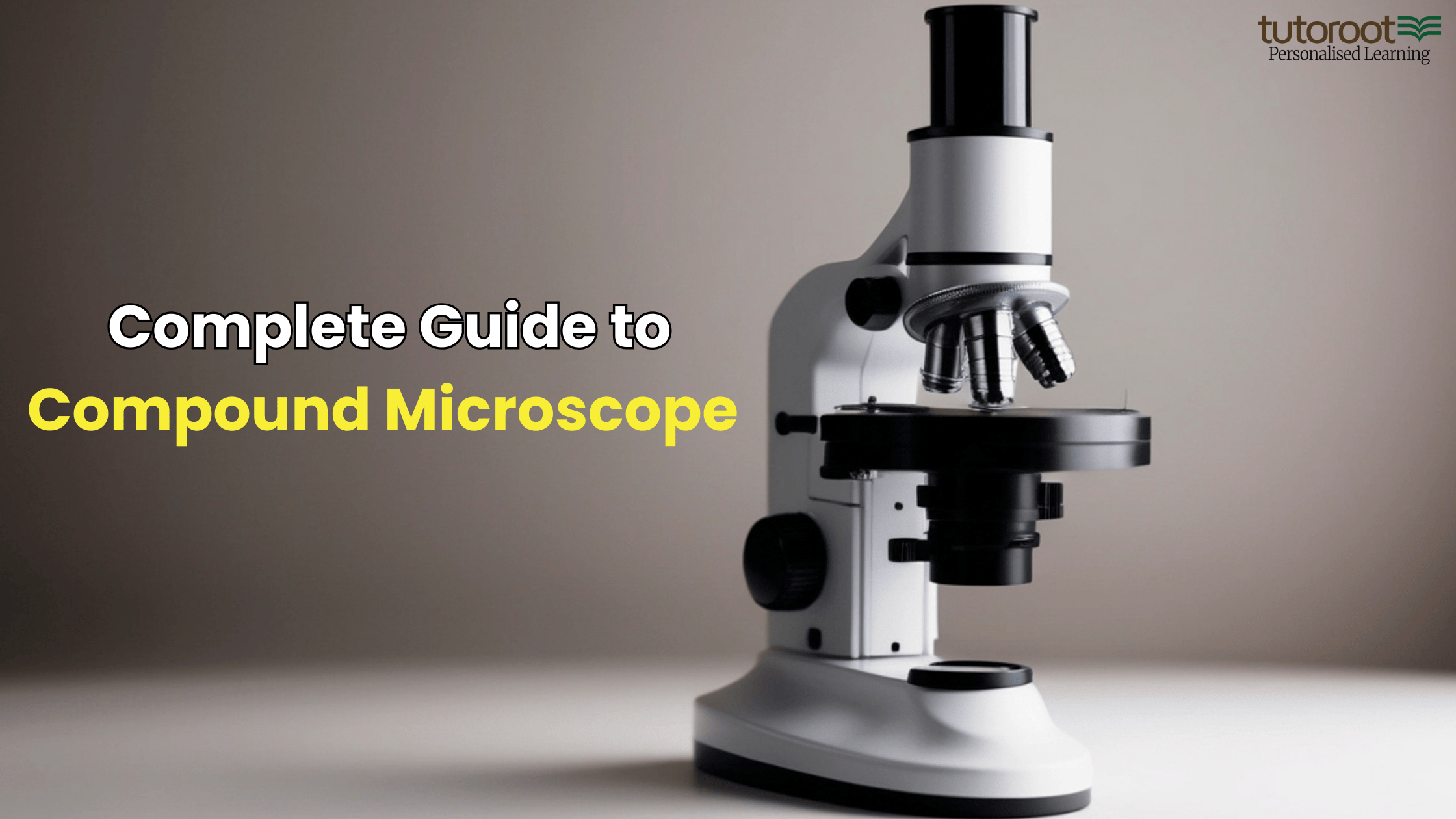
Compound Microscope Diagram
The compound microscope diagram shows the arrangement of optical and non-optical parts such as:
- Eyepiece
- Objective lenses
- Nosepiece
- Stage
- Mirror / light source
- Coarse and fine adjustment knobs
📌 Diagram-based questions from the compound microscope are very common in board and NEET exams.
Compound Microscope Ray Diagram
The compound microscope ray diagram explains how light passes through:
-
The transparent specimen
-
Objective lens (forms real, inverted image)
-
Ocular lens (forms final virtual image)
This ray diagram helps students understand:
- Image formation
- Magnification concept
- Bright-field nature of microscope
Working Principle of Compound Microscope
The working principle of a compound microscope is based on refraction of light through lenses.
How it Works:
- The objective lens, placed close to the object, forms a real and magnified image
- This image acts as an object for the ocular lens
- The ocular lens further magnifies it to form a virtual image
- Light travels directly from the source to the eye, making it a bright-field microscope
Parts of Compound Microscope
The compound microscope parts are classified into:
1. Non-Optical Parts
- Base – Supports the microscope
- Pillar – Connects base and arm
- Arm – Holds stage and body tube
- Inclination Joint – Allows tilting
- Stage – Platform to place slide
- Body Tube – Holds lenses
- Rack and Pinion – Used for focusing
- Coarse & Fine Adjustment Screws – Sharpens image
2. Optical Parts
- Diaphragm – Controls light intensity
- Condenser – Focuses light on specimen
- Mirror (Reflector) – Directs light
- Objective Lenses – Low power, high power, oil immersion
- Ocular Lens (Eyepiece) – 5X, 10X, 15X, 20X
These compound microscope parts and functions are frequently asked in exams.
Compound Microscope Drawing (For Exams)
In exams, students are often asked to draw and label a compound microscope.
While making a compound microscope drawing, ensure:
-
Proper labeling
-
Neat structure
-
Correct position of lenses and stage
📌 Practice diagram-based questions to score full marks.
Advantages and Disadvantages of Compound Microscope
Advantages
- Provides clear and detailed 2D images
- Built-in light source
- Ideal for school and laboratory use
Disadvantages
- Limited magnification compared to electron microscopes
- Only transparent specimens can be observed
Conclusion
The compound microscope is a fundamental topic in Physics and Biology. Knowing its diagram, ray diagram, parts, working principle, and drawing helps students score better in board exams and NEET.
Learn Physics Better with Tutoroot 🌱

At Tutoroot, we simplify complex topics like the compound microscope using:
-
Expert Physics faculty
-
Concept clarity with diagrams
-
Exam-focused preparation
-
Regular doubt-clearing sessions
🎯 Start with a FREE DEMO class today and experience personalised learning that builds confidence and results.
👉 Explore more Physics blogs on Tutoroot for simplified explanations and smart learning strategies.
FAQs on Compound Microscope
1. What is a compound microscope?
A compound microscope is an optical instrument that uses two lenses to magnify small objects.
2. What is shown in a compound microscope diagram?
It shows the labeled optical and non-optical parts of the microscope.
3. What is a compound microscope ray diagram?
It explains image formation through objective and ocular lenses.
4. What are the main parts of a compound microscope?
Objective lens, eyepiece, stage, mirror, condenser, and adjustment knobs.
5. Why is compound microscope important for students?
It is a core topic in Class 9–12 and NEET exams.
Visit Tutoroot Social Platforms:
![]()
![]()
![]()
![]()
![]()
Everything is very open with a precise explanation of
the issues. It was truly informative. Your site is very helpful.
Thank you for sharing!
Great article, totally what I needed.
It’s remarkable in support of me to have
a site, which is valuable for my knowledge. thanks admin
My relatives every time say that I am killing my time here
at web, however I know I am getting experience every day by reading such
nice articles
An intriguing discussion is definitely worth comment.
I do believe that you should publish more about this subject, it may not be a taboo matter but
usually folks don’t speak about such subjects. To the next!
Cheers!!
Good artcile, but it would be better if in future you
can share more about this subject. Keep posting.