
Human Heart – Functions, Circulation, Diagram
The human heart is a muscular fist-sized organ that is situated in the front of the chest to pump blood throughout the body. The human heart is divided into four major portions (chambers) that are driven by electrical impulses. Its function is controlled by the brain and the neurological system.
The human heart is one of the most important organs in charge of keeping life going. It is a four-chambered muscular organ. The human heart is one of the strongest and most hardworking muscles in the human body, working continuously throughout a person’s life.
Aside from humans, most other animals have a heart that circulates blood throughout their bodies. Even invertebrates, such as grasshoppers, have a pumping organ like the human heart, though it does not operate in the same manner.
In this article, we shall learn more about the structure and functions of the human heart.
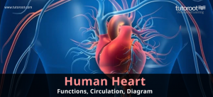
The human heart diagram, to help understand better the structure of the human heart, has also been provided for your benefit
Structure of the Human Heart
Let us get into the basic anatomy of the human heart.
The human heart is positioned in the thoracic cavity between the lungs, somewhat to the left of the sternum (breastbone). It is around the size of a fist and is split into four chambers, two ventricles, and two atria. The ventricles are the blood-pumping chambers, whereas the atrium is the blood-receiving chamber. The right atrium and ventricle comprise the “right heart,” whereas the left atrium and ventricle comprise the “left heart.” The heart anatomy also houses the largest artery in the body, the aorta. The chambers of the heart are driven by electrical impulses.
The human heart is located between the lungs and is behind the sternum and above the diaphragm. A sac called the pericardium surrounds the heart. As explained, two of the chambers of the human heart are located on the top, while the other two are on the bottom.
Let us learn more about the structure of the human heart by understanding the anatomy and functions of its individual parts:
Heart Chambers
The number of chambers of a vertebrate heart can be used to classify it. Fish, for example, has two chambers, but reptiles and amphibians have three chambers. Whereas birds and mammals have 4 chambers. Humans also have four chambers:
- The left atrium
- The right atrium
- The left ventricle
- The right ventricle
As mentioned above, two chambers, the right ventricle and the left ventricle of the human heart are the bottom chambers and are separated by a wall named as the interventricular septum.
The top chambers in the structure of the human heart are the right and left atria which are separated by a wall called the interatrial septum.
Atria
Atria are thinner and have less muscular walls than ventricles. These are the blood-receiving chambers that are connected by large veins.
Ventricles
Ventricles or the lower chambers of the human heart are bigger and more muscular chambers that pump and push blood into circulation. These are linked to bigger arteries, which transport blood for circulation.
If you notice the structure of the heart vis-à-vis its chambers, it is clearly understood that the right ventricle and atrium are much smaller than the left chambers. The walls have fewer muscles than the left side, and the size difference is due to their roles. Blood from the right side goes through pulmonary circulation, whereas blood from the left side is circulated throughout the body. Please check the human heart diagram for a more effective understanding through illustration.
The functions of the human heart are thus primarily determined by the proper state of its chambers.
Blood Vessels
Another important component that drives the proper and effective functions of the human heart is its blood vessels. Blood travels via channels of varied diameters in creatures with closed circulatory systems. Such type of circulation is seen in all animals, including humans. Many blood veins create a network on the heart’s exterior surface, with additional significant vessels originating from within the tissue.
Blood vessels are tube-like structures and consist of three components – veins, arteries, and capillaries. In order to differentiate the shape, size, and functioning of these components, one must analyze aspects such as their location, oxygen content in the blood, valves’ and muscle tissues’ presence, the direction in which the blood travels, and of course the size of the wall.
Veins
Veins provide deoxygenated blood to the heart through the inferior and superior vena cava, where it empties into the right atrium. The veins are thin walls and are in the periphery of the skin and much closer to it. Unlike arteries, they are not muscular and depend on the valves for the flow of blood. From tiny blood vessels called venules, these veins elongate into full-sized vessels when they are closer to the human heart.
Capillaries
Capillaries are small, tube-like that connect the arteries and veins. They typically carry blood between the other two blood vessel categories. Capillaries are the smallest among the blood vessels, as little as 5 micrometers, even less than the width of human hair. The wall of a capillary is one cell as far as its thickness is concerned. It is made of endothelial cells. A capillary lets oxygen, wastes, and even nutrients to pass through tissue cells. Capillaries do not have valves or muscle tissues. They can carry both oxygenated and deoxygenated blood.
Arteries
Arteries are muscular-walled tubes that transport oxygenated blood from the heart to all other regions of the body. Arteries are made up of thick walls and a strong layer that is muscular in its make and facilitates the flow of blood. The arteries are the largest and the widest of the blood vessels.
The Arteries are made up of further smaller units called arterioles. These along with primary arteries alter their size in order to maintain the levels of blood pressure. The aorta is the major artery and branches into several smaller arteries throughout the body. It carries blood from the heart to organs. Arteries are located very deep inside the muscle. They have no valves present within, with the pulmonary artery being the exception.
While understanding the functions of the heart, it is vital to learn about the differences in the functioning of blood vessels.
Valves
Valves are fibrous tissue flaps located between the veins in the heart chambers. They make certain that blood flows in only one direction (unidirectional). The flaps retain blood from flowing backward. Valves separate the top and bottom chambers of the human heart and carry blood too.
Valves are classified according to their function. They are:
Atrioventricular valves
Atrioventricular valves exist between the ventricles and the atria. The tricuspid valve connects the right ventricle to the right atrium, whereas the mitral valve connects the left ventricle to the left atrium.
Semilunar valves
Semilunar valves exist between the left ventricle and the aorta. It is also near the right ventricle and the pulmonary artery.
Heart Walls
The heart walls are essential vital in pumping blood against strong resistance due to the arteries. The blood that is disposed of inside the left ventricle needs high pressure so that it is smoothly transferred to the rest of the body. This makes it essential for the heart walls to be substantially thick. The heart walls have three major layers:
Epicardium
The epicardium is the heart’s outermost layer. It is made up of a thin-layered membrane that lubricates and protects the outer part. It is the visceral layer of the serous pericardium, which adheres to the heart’s myocardium. In terms of histology, epicardium is made of mesothelial cells.
Myocardium
This is a layer of muscle tissue that makes up the middle layer of the human heart’s wall. It adds to the thickness and oversees the pumping motion. In terms of its functions, the myocardium is the main part of the heart and is the thickest of three layers. Being a muscle layer, it enables contractions of the heart. As far of histology is concerned, the myocardium is made up of cardiomyocytes that contain rich reserves of glycogen and mitochondria.
Endocardium
The endocardium is the deepest or innermost layer of the human heart that borders the inner chambers and covers the heart valves. Furthermore, it keeps blood from adhering to the inner walls of the vessels, preventing potentially dangerous blood clots. It is made of two layers, and the inner one is made up of endothelial cells, like epicardium.
Functions of the Human Heart
In every organism, the job of the heart is to maintain blood flowing constantly throughout the body. This replaces the oxygen supply and distributes nutrients throughout the cells and tissues.

The heart’s primary functions are as follows:
- The human heart’s duty is to circulate blood throughout the body.
- Throughout the body, including the human heart, blood delivers oxygen, hormones, glucose, and other chemicals.
- The heart also keeps the blood pressure in the body steady.
There are two forms of circulation within the body: pulmonary circulation and systemic circulation. And these are the important function of Human Heart.
Human Heart Blood Circulation
Pulmonary circulation
Pulmonary circulation is the component of circulation that transports deoxygenated blood from the heart to the lungs and subsequently returns oxygenated blood to the heart.
Systemic circulation
This is another kind of circulation is systemic circulation, in which oxygenated blood is pumped from the heart to every organ and tissue in the body, and deoxygenated blood returns to the heart.
As the heart is also a muscle, even it requires a steady supply of oxygenated blood. This is when another sort of circulation, the coronary circulation, comes into action.
Coronary circulation
Coronary circulation is a vital component of circulation that supplies oxygenated blood to the heart. This is significant since the heart is in charge of delivering blood throughout the body.
Final Notes
We hope this article clears all your doubts about Human heart, as well we covered in-depth the function of human heart, and human heart diagram.
Tutoroot Biology lessons can help you learn more about the construction and function of the human heart, as well as other relevant concepts. A personalized tutoring mentor can assist you in learning more about such Biology subjects.
FAQ’s
What is the position of the human heart in the body?
The human heart is located in the chest where the sternum sits behind and slightly to the left of your breastbone.
What is the normal heart rate of a human?
A normal resting heart rate should be 72 beats per minute, but it can vary from 60-100 beats.
How many chambers are there in the human heart?
There are 4 chambers in a typical heart, two upper and two lower chambers.
What are the 4 main parts of the human heart?
The four main parts of the human heart are chambers, blood vessels, valves, and walls
Which side is heart pain?
Generally, heart pain happens on the left side or towards the center of the chest
What is the primary function of the human heart?
The primary function of the human heart is to circulate blood throughout the body.